Wednesday 28 December 2011

Water. How trying to lose weight is helping me to rediscover me.

Current weight:- 14 stones.

 Target weight: 10 stones
Time span to achieve goal: 6 months.
So I'll have to drink loads of water.

More water,
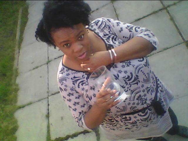
                                         and even more water.

I am starting to realise how beautiful I am and I remember how great it feels as my weight is starting to come off.

I have to plan all my meals now; which are mainly col or haddock (steamed with garlic) and prepacked seasonal salads.

Weightloss is very important to self-esteem and I have found out that, it does not have to be miserable.  It really can be fun, especially when you are sharing your journey with the entire world..

Tuesday 27 December 2011

Drink water when you feel hungry...


I am going to be disciplined and do proper training and eat nutritious foods in small amounts to achieve a toned body.


Water rehydrates the cells.....


 No need to spend money on pills or anything like that. Only my willpower and discipline is what I am going to use to get a natural toned body.


I have increased my consumption of water and cut out all alcohol..


Also I try to eat Broccoli with all meals.


Monday 26 December 2011

An Introduction to Mind, Body and Soul (WeightLoss)

Hi friends & followers of natural.Gift,


Mind, body and soul, weight loss...


I'm just using this initial post, to introduce myself, and let you know what my goals & aspirations are for this weight loss blog.  


My start weight is 13 1/2 stones.

 I am a teacher by profession, but I am involved in so many more aspects of life, for instance, I am a wife and mother of six beautiful children.  They are so wonderful, they look up to me, whatever I do; three years ago, one of my children died of a brain tumor, Josh, so he is my main reason, for getting up off of my backside, and do something.......  He was 15 years old...all I remember him saying is, "I'm not afraid to die, mom!".  Why do we even have to die at all, who says so.......  So, he is my inspiration, to win this battle........


My main ethos is that only you can change your situation.  You are the g.o.d. (giver of desire) of your world, and if you want to make a difference, in your life;  you have to get up and just do it....All our thoughts are part of the morphogenic field, all our thoughts matter, they are interlinked and inter connected..... Our thoughts creates atoms, they are entities all by their selves, so at all times, I believe I should think positively...


About 10 years ago, I was diagnosed with a chronic, autoimmune illness called Sarcoidosis; with which Bernie Mac & others have lived with & died from.....It effects every single organ in my body; my eyes, my skin, my lungs, my heart, my liver,  


I was prescribed the palliative treatment, of steroids, Prednisolone which will never cure me of this dreaded illness, but it helps to alleviate the symptoms.... But unfortunately, it causes weight gain, water retention, high blood pressure, etc..........


Here comes the science bit


Sarcoidosis (from sarc meaning flesh, -oid, like, and -osis, diseased or abnormal condition), also called sarcoid, Besnier-Boeck disease or Besnier-Boeck-Schaumann disease, is a disease in which abnormal collections of chronic inflammatory cells (granulomas) form as nodules in multiple organs.[1] The cause of sarcoidosis is unknown. Granulomas most often appear in the lungs or the lymph nodes, but virtually any organ can be affected. Normally the onset is gradual. Sarcoidosis may be asymptomatic or chronic. It commonly improves or clears up spontaneously. More than 2/3 of people with lung sarcoidosis have no symptoms after 9 years. About 50% have relapses. About 10% develop serious disability. Lung scarring or infection may lead to respiratory failure and death.[1] Chronic patients may deal with waxing and waning symptoms over many years.....


I used to be maybe nine stones 10 years ago, now I am 141/2 stones with high blood pressure....So I'm making a proactive effort to alkalize my mind, body and soul and heal myself............


Sarcoidosis

From Wikipedia, the free encyclopedia
Jump to: navigation, search
Sarcoidosis
Classification and external resources

Chest xray showing the typical nodularity of sarcoidosis in the base of the lungs.
ICD-10D86
ICD-9135
OMIM181000
DiseasesDB11797
MedlinePlus000076
eMedicinemed/2063
MeSHD012507
Sarcoidosis (from sarc meaning flesh, -oid, like, and -osis, diseased or abnormal condition), also called sarcoid, Besnier-Boeck disease or Besnier-Boeck-Schaumann disease, is a disease in which abnormal collections of chronic inflammatory cells (granulomas) form as nodules in multiple organs.[1] The cause of sarcoidosis is unknown. Granulomas most often appear in the lungs or the lymph nodes, but virtually any organ can be affected. Normally the onset is gradual. Sarcoidosis may be asymptomatic or chronic. It commonly improves or clears up spontaneously. More than 2/3 of people with lung sarcoidosis have no symptoms after 9 years. About 50% have relapses. About 10% develop serious disability. Lung scarring or infection may lead to respiratory failure and death.[1] Chronic patients may deal with waxing and waning symptoms over many years.[2]


Signs and symptoms

Signs and symptoms of sarcoidosis.[3]
Sarcoidosis is a systemic disease that can affect any organ. Common symptoms are vague, such as fatigue unchanged by sleep, lack of energy, weight loss, aches and pains, arthritis, dry eyes, swelling of the knees, blurry vision, shortness of breath, a dry hacking cough or skin lesions. Sarcoidosis and cancer may mimic one another, making the distinction difficult.[4] The cutaneous symptoms vary, and range from rashes and noduli (small bumps) to erythema nodosum or lupus pernio. It is often asymptomatic.
The combination of erythema nodosum, bilateral hilar lymphadenopathy and arthralgia is called Löfgren syndrome. This syndrome has a relatively good prognosis.
Renal, liver (including portal hypertension), heart[5] or brain involvement may cause further symptoms and altered functioning.

Lungs

Of individuals with sarcoidosis, 90 percent have an abnormal chest x-ray at some time during their course. Overall, approximately 50 percent develop permanent pulmonary abnormalities and 5 to 15 percent have progressive fibrosis of the lung parenchyma. Sarcoidosis of the lung is primarily an interstitial lung disease in which the inflammatory process involves the alveoli, small bronchi, and small blood vessels. In acute and subacute cases, the physical examination usually reveals dry rales.[6]

Liver

Although liver biopsy reveals liver involvement in 60 to 90 percent of cases, liver dysfunction is usually not important clinically. Approximately 20–30% have hepatomegaly and/or biochemical evidence of liver involvement. Usually these changes reflect a cholestatic pattern and include an elevated alkaline phosphatase level; the bilirubin and aminotransferases are only mildly elevated. Jaundice is rare.[6]

Skin

Sarcoidosis involves the skin in about 25 percent of patients. The most common lesions are erythema nodosum, plaques, maculopapular eruptions, subcutaneous nodules, and lupus pernio. Treatment is not required, since the lesions usually resolve spontaneously in 2 to 4 weeks. Although it may be disfiguring, cutaneous sarcoidosis rarely causes major problems.[6]

Heart

Although cardiac involvement is present in 20% to 30% of patients with sarcoidosis, only about 5% of patients with systemic sarcoidosis are symptomatic.[7]
The presentation of cardiac sarcoidosis can range from asymptomatic conduction abnormalities to fatal ventricular arrhythmia.[8] Myocardial sarcoidosis can be a rare cause of sudden cardiac death.[9][10]

Eye

Manifestations in the eye include uveitis, uveoparotitis, and retinal inflammation, which may result in loss of visual acuity or blindness. The combination of anterior uveitis, parotitis, VII cranial nerve paralysis and fever is called uveoparotid fever, and is associated with Heerfordt-Waldenstrom syndrome. (D86.8)

Blood

Abnormal clinical blood tests are frequent but not diagnostic. Anemia occurs in 4-20% of patients with sarcoidosis. Leukopenia (due to a reduced number of circulating lymphocytes [11] or lymphopenia) occurs in as many as 40% of patients but is rarely severe. In the absence of splenomegaly, leukopenia may reflect bone marrow involvement, however, the most common mechanism is a redistribution of blood T cells to sites of disease.[12] Other non-specific findings include monocytosis, occurring in the majority of sarcoidosis cases,[13] increased hepatic enzymes or alkaline phosphatase. Hypercalciuria and hypercalcemia are seen in <10% of patients.[14]


Lymph nodes
Lymphadenopathy is very common in sarcoidosis. Intrathoracic nodes are enlarged in 75 to 90 percent of all patients; usually this involves the hilar nodes, but the paratracheal nodes are commonly involved. Peripheral lymphadenopathy is very common, particularly involving the cervical (the most common head and neck manifestation of the disease [15]), axillary, epitrochlear, and inguinal nodes. Palpation causes no pain.[6]


 Nervous system
All components of the nervous system can be involved in sarcoidosis. Sarcoidosis affecting the brain or nerves is known as neurosarcoidosis. Neurologic findings are observed in about 5 percent of patients. Seventh nerve involvement with unilateral facial paralysis is most common. It occurs suddenly and is usually transient. Other common manifestations of neurosarcoid include optic nerve dysfunction, papilledema, palate dysfunction, hearing abnormalities, hypothalamic and pituitary abnormalities, chronic meningitis, and peripheral neuropathy.[6] Intramedullary sarcoidosis is rare and occurs in less than 1% of cases. There is usually granulomatous involvement of the basal meninges that subsequently affects the cranial nerves. Myelopathy may be the initial clinical presentation of intramedullary neurosarcoidosis.[16]

Exocrine glands

Parotid enlargement is a classic feature of sarcoidosis, but clinically apparent parotid involvement occurs in less than 10 percent of patients. Bilateral involvement is the rule. The gland is usually nontender, firm, and smooth. Xerostomia can occur; other exocrine glands are affected only rarely.[6]

Scalp

Sarcoidosis of the scalp presents with diffuse or patchy hair loss.[17]:762

Causes

The exact cause of sarcoidosis is not known. The current working hypothesis is that in genetically susceptible individuals sarcoidosis is caused through alteration in immune response after exposure to an environmental, occupational, or infectious agent.[18]

Genetics

Investigations of genetic susceptibility yielded many candidate genes but only few were confirmed by further investigations and no reliable genetic markers are known. Currently, the most interesting candidate gene is BTNL2; several HLA-DR risk alleles are also being investigated.[19] In persistent sarcoidosis the HLA haplotype HLA-B7-DR15 are either cooperating in disease or another gene between these two loci is associated. In non-persistent disease there is a strong genetic association with HLA DR3-DQ2.[20] Siblings have only a modestly increased risk (hazard ratio 5-6) of developing the disease, indicating that genetic susceptibility plays only a small role. The alternate hypothesis that family members share similar exposures to environmental pathogens is quite plausible to explain the apparent hereditary factor.

Infectious agents

Several infectious agents appear to be significantly associated with sarcoidosis but none of the known associations is specific enough to suggest a direct causative role. Propionibacterium acnes can be found in bronchoalveolar lavage of approximately 70% patients and is associated with disease activity, however it can be also found in 23% of controls.[21][22] A recent meta-analysis investigating the role of mycobacteria in sarcoidosis found it was present in 26.4% of cases, however the meta-analysis also detected a possible publication bias, so the results need further confirmation.[23][24]
There have also been reports of transmission of sarcoidosis via organ transplants.[25]

Vitamin D dysregulation

Sarcoidosis frequently causes an increase in vitamin D production outside the kidney.[26] Macrophages inside the granulomas convert vitamin D to its active form, resulting in elevated levels of the hormone 1,25-dihydroxyvitamin D and symptoms of hypervitaminosis D that may include fatigue, lack of strength or energy, irritability, metallic taste, temporary memory loss or cognitive problems. Physiological compensatory responses (e.g., suppression of the parathyroid hormone levels) may mean the patient does not develop frank hypercalcemia. This condition may be aggravated by high levels of estradiol and prolactin such as in pregnancy, leading to hypercalciuria and/or compensatory hypoparathyroidism.[27] High levels of Vitamin D are also implicated in immune-system dysfunctions which tie into the sarcoid condition.

Hyperprolactinemia

Prolactin is frequently increased in sarcoidosis, between 3–32% cases have hyperprolactinemia,[28] this frequently leads to amenorrhea, galactorrhea or nonpuerperal mastitis in women. Prolactin also has a broad spectrum of effects on the immune system and increased prolactin levels are associated with disease activity or may exacerbate symptoms in many autoimmune diseases and treatment with prolactin lowering medication has been shown effective in some cases.[29] However it is unknown if this relation holds in sarcoidosis and the gender predilection in sarcoidosis is less pronounced than in some other autoimmune diseases where such relation has been established. In pregnancy, the effects of prolactin and estrogen counteract each other to some degree, with a slight trend to improve pulmonary manifestations of sarcoidosis while lupus, uveitis and arthralgia might slightly worsen.[27] Lupus, uveitis and arthralgia are known to be in some cases associated with increased prolactin levels and respond to bromocriptin treatment but so far this has not been investigated specifically for sarcoidosis. The reasons for increased prolactin levels in sarcoidosis are uncertain. It has been observed that prolactin is produced by T-lymphocytes in some autoimmune disorders in amounts high enough to affect the feedback by the hypothalamic dopaminergic system.[30]
The extrapituitary prolactin is believed to play a role as a cytokine like proinflammatory factor. Prolactin antibodies are believed to play a role in hyperprolactinemia in other autoimmune disorders and high prevalence endocrine autoimmunity has been observed in patients with sarcoidosis.[31] It may also be a consequence of renal disease or treatment with steroids. Neurosarcoidosis may occasionally cause hypopituiarism but has not been reported to cause hyperprolactinemia.

Thyroid disease

In women, a substantial association of thyroid disease and sarcoidosis has been reported. The association is less marked but still significant for male patients. Female patients have a significantly elevated risk for hypothyroidism, hyperthyroidism and thyroid autoimmunity and it appears that autoimmunity is very important in the pathogenesis of thyroid disease in this population. Thyroid granulomatosis on the other hand is uncommon.[32]

Autoimmune

Association of autoimmune disorders has been frequently observed. The exact mechanism of this relation is not known but some evidence supports the hypothesis that this is a consequence of Th1 lymphokine prevalence.[32][33]
Sarcoidosis has been associated with celiac disease. Celiac disease is a condition in which there is a chronic reaction to certain protein chains, commonly referred to as glutens, found in some cereal grains. This reaction causes destruction of the villi in the small intestine, with resulting malabsorption of nutrients.
An association with type IV hypersensitivity has been described.[34] Tests of delayed cutaneous hypersensitivity have been used to measure progression.[35]

Other

While disputed, some cases have been associated with inhalation of the dust from the collapse of the World Trade Center after the September 11, 2001 attacks.[36] See Health effects arising from the September 11, 2001 attacks for more information. Chicago comedian, Bernie Mac, suffered from sarcoidosis and died of pneumonia as a result of his compromised immune system.[37] Reggie White, a former standout National Football League player, also suffered from sarcoidosis, and the disease played a major role in his death.[38]

Pathophysiology

Granulomatous inflammation is characterized primarily by accumulation of monocytes, macrophages and activated T-lymphocytes, with increased production of key inflammatory mediators, TNF-alpha, IFN-gamma, and IL-12, characteristic of a Th1-polarized response (T-helper lymphocyte-1 response). Sarcoidosis has paradoxical effects on inflammatory processes; it is characterized by increased macrophage and CD4 helper T-cell activation resulting in accelerated inflammation, however, immune response to antigen challenges such as tuberculin is suppressed. This paradoxic state of simultaneous hyper- and hypo- activity is suggestive of a state of anergy. The anergy may also be responsible for the increased risk of infections and cancer. It appears that regulatory T-lymphocytes in the periphery of sarcoid granulomas suppress IL-2 secretion which is hypothesized to cause the state of anergy by preventing antigen-specific memory responses.[39]
While it is widely believed that TNF-alpha plays an important role in the formation of granulomas, it was observed that sarcoidosis can be triggered by treatment with the TNF-alpha antagonist etanercept.[40][41]

Diagnosis

Diagnosis of sarcoidosis is often a matter of exclusion. To exclude sarcoidosis in a case presenting with pulmonary symptoms might involve chest X-ray, CT scan of chest, PET scan, CT-guided biopsy, mediastinoscopy, open lung biopsy, bronchoscopy with biopsy, endobronchial ultrasound and endoscopic ultrasound with FNA of mediastinal lymph nodes(EBUS FNA). Tissue from biopsy of lymph nodes is subjected to both flow cytometry to rule out cancer and special stains (acid fast bacilli stain and Gömöri methenamine silver stain) to rule out microorganisms and fungi. Angiotensin-converting enzyme blood levels are used in diagnosis and monitoring of sarcoidosis.[42]
Differential diagnosis includes metastatic disease, lymphoma, septic emboli, rheumatoid nodules, Wegener's granulomatosis, varicella infection, and atypical infections such as mycobacterium avium complex, cytomegalovirus, and cryptococcus.[43] Sarcoidosis is confused most commonly with neoplastic diseases such as lymphoma or with disorders characterized also by a mononuclear cell granulomatous inflammatory process, such as the mycobacterial and fungal disorders.[6]
Because of the wide range of possible manifestations the investigations to confirm diagnosis may involve many organs and methods depending on initial presentation.
Very often, Sarcoidosis presents as a restrictive disease of the lungs, causing a decrease in lung volume and decreased compliance (the ability to stretch) — hence chest X-ray and other methods are used to assess the severity or rule out pulmonary disease.
The disease typically limits the amount of air drawn into the lungs, but produces higher than normal expiratory flow ratios. The vital capacity (full breath in, to full breath out) is decreased, and most of this air can be blown out in the first second. This means the FEV1/FVC ratio is increased from the normal of about 80%, to 90%. Obstructive lung changes, causing a decrease in the amount of air that can be exhaled, may occur when enlarged lymph nodes in the chest compress airways or when internal inflammation or nodules impede airflow.

CT scan of the chest showing lymphadenopathy (arrows) in the mediastinum due to sarcoidosis.
Chest X-ray changes are divided into four stages
  • Stage 1 bihilar lymphadenopathy
  • Stage 2 bihilar lymphadenopathy and reticulonodular infiltrates
  • Stage 3 bilateral pulmonary infiltrates
  • Stage 4 fibrocystic sarcoidosis typically with upward hilar retraction, cystic & bullous changes
Although patients with type I x-rays tend to have the acute or subacute, reversible form of the disease while those with types II and III often have the chronic, progressive disease, these patterns do not represent consecutive "stages" of sarcoidosis. Thus, except for epidemiologic purposes, this x-ray categorization is mostly of historic interest.[6]
Investigations to assess involvement of other organs frequently involve electrocardiogram, ocular examination by an ophthalmologist, liver function tests, renal function tests, serum calcium and 24-hour urine calcium.
In Sarcoidosis presenting in the Caucasian population, hilar adenopathy and erythema nodosum are the most common initial symptoms. In this population, a biopsy of the gastrocnemius muscle is a useful tool in correctly diagnosing the patient. The presence of a noncaseating epithelioid granuloma in a gastrocnemius specimen is definitive evidence of sarcoidosis as other tuberculoid and fungal diseases extremely rarely present histologically in this muscle[44].
In female patients, sarcoidosis is significantly associated with hypothyroidism, hyperthyroidism and other thyroid diseases, hence close surveillance of thyroid function is recommended [32]

[edit] Classification

Sarcoidosis may be divided into the following types:[17]:708-11

Treatment

Between 30 and 70% of patients do not require therapy.[45] For patients presenting with lung symptoms, unless the respiratory impairment is devastating, active pulmonary sarcoidosis is observed usually without therapy for 2 to 3 months; if the inflammation does not subside spontaneously, therapy is instituted.[6] Corticosteroids, most commonly prednisolone, have been the standard treatment for many years. In some patients, this treatment can slow or reverse the course of the disease, but other patients do not respond to steroid therapy. The use of corticosteroids in mild disease is controversial because in many cases the disease remits spontaneously.[46] Additionally, corticosteroids have many recognized dose- and duration-related side effects, and their use is generally limited to severe, progressive, or organ-threatening disease. The influence of corticosteroids or other immunosuppressants on the natural history is unclear.
Severe symptoms are generally treated with steroids, and steroid-sparing agents such as azathioprine and methotrexate are often used. Rarely, cyclophosphamide has also been used. As the granulomas are caused by collections of immune system cells, particularly T cells, there has been some early indications of success using immunosuppressants, interleukin-2 inhibitors or anti-tumor necrosis factor-alpha treatment (such as infliximab). Unfortunately, none of these has provided reliable treatment, and there can be significant side effects such as an increased risk of reactivating latent tuberculosis. Anti-tumor necrosis factor-alpha treatment with etanercept in rheumatoid arthritis has been observed to cause sarcoidosis.[40]
Because sarcoidosis can affect multiple organ systems, follow-up on a patient with sarcoidosis should always include an electrocardiogram, ocular examination by an ophthalmologist, liver function tests, serum calcium and 24-hour urine calcium. In female patients sarcoidosis is significantly associated with hypothyroidism, hyperthyroidism and other thyroid diseases, hence close surveillance of thyroid function is recommended.[32]

Prognosis

The disease can remit spontaneously or become chronic, with exacerbations and remissions. In some patients, it can progress to pulmonary fibrosis and death. Approximately half of the cases resolve without treatment or can be cured within 12–36 months and most within 5 years. Some cases persist several decades.[45] Where the heart is involved, the prognosis is poor.[47] Patients with sarcoidosis appear to be at significantly increased risk for cancer, in particular lung cancer, malignant lymphomas,[48] and cancer in other organs known to be affected in sarcoidosis.[49] In sarcoidosis-lymphoma syndrome, sarcoidosis is followed by the development of a lymphoproliferative disorder such as non-Hodgkin lymphoma.[50] This may be attributed to the underlying immunological abnormalities that occur during the sarcoidosis disease process.[51] Sarcoidosis can also follow cancer [52] or occur concurrently with cancer.[53][54] There have been reports of hairy cell leukemia,[55] acute myeloid leukemia,[56] and acute myeloblastic leukemia [57] associated with sarcoidosis.

Epidemiology

Sarcoidosis most commonly affects young adults of both sexes, although studies have reported more cases in females. Incidence is highest for individuals younger than 40 and peaks in the age-group from 20 to 29 years; a second peak is observed for women over 50.[45][47]
Sarcoidosis occurs throughout the world in all races with an average incidence of 16.5/100,000 in men and 19/100,000 in women. The disease is most prevalent in Northern European countries, and the highest annual incidence of 60/100,000 is found in Sweden and Iceland. In the United States, sarcoidosis is more common in people of African descent than Caucasians, with annual incidence reported as 35.5 and 10.9/100,000, respectively.[58] Sarcoidosis is less commonly reported in South America, Spain, India, Canada, and the Philippines.There may be a higher susceptibility to sarcoidosis in those with coeliac disease. An association between the two disorders has been suggested.[59]
The differing incidence across the world may be at least partially attributable to the lack of screening programs in certain regions of the world and the overshadowing presence of other granulomatous diseases, such as tuberculosis, that may interfere with the diagnosis of sarcoidosis where they are prevalent.[47] There may also be differences in the severity of the disease between people of different ethnicities. Several studies suggest that the presentation in people of African origin may be more severe and disseminated than for Caucasians, who are more likely to have asymptomatic disease.
Manifestation appears to be slightly different according to race and sex. Erythema nodosum is far more common in men than in women and in Caucasians than in other races. In Japanese patients, ophthalmologic and cardiac involvement are more common than in other races.
Sarcoidosis is one of the few pulmonary diseases with a higher prevalence in non-smokers.

In pregnancy

Sarcoidosis generally does not prevent successful pregnancy and delivery; the endogenous estrogen in pregnancy may even have a slightly beneficial immunomodulatory effect. In most cases the course of sarcoidosis is unaffected by pregnancy; there is improvement in a few cases and worsening of symptoms in very few cases.....


So basically, that's the reason; I've started this blog, to share and encourage others on my journey to divine healing and ultimate health and well being...........


I hate using the steriods, the side effects are devastating.  My next hospital appointment is January 2011, I have to have x-rays to look at my lungs, they take my blood and weight me....I know my B.M.I.  is about 33, so that is something I will be actively trying to reduce....


An overview of steroid use and its potential side-effects
Steroids are fatty or lipid compounds formed by the body from cholesterol which is produced in the liver and used by the adrenal glands in the synthesis of these hormones. In everyday practice patients are prescribed steroids to treat a range of conditions from asthma through arthritis, eczema, multiple sclerosis, leukaemia and many other diseases (plus of course COP/BOOP). Pharmacologically they are used in much larger doses: their action is twofold, to suppress inflammation and the immune response. Steroids lower white cell counts and antibody formation, immunosuppression with prednisolone occurs at doses in excess of 20 mg per day. The immune response is suppressed to prevent organ transplant rejection or to treat severe systemic allergic reactions. Health professionals are likely to watch out for the side-effects of the immune and inflammatory suppression.
below: chemical structure of prednisone (left) and prednisolone (right) which are very similar compounds
Steroids are classified as either short-to-medium acting, intermediate or long acting. Short acting steroids include hydrocortisone, cortisone, prednisone (an inactive form converted to prednisolone in the liver) and prednisolone.
Effects on Adrenal Cortex
The administration of steroids for a period exceeding two weeks runs the risk of suppression of the adrenal cortex. Patients on steroids for less than one week may have them stopped without the risk of adrenal suppression. In people who take large doses of corticosteroids, such as prednisone or prednisolone the function of the adrenal glands will become suppressed. This suppression occurs because these large doses prevent the hypothalamus and pituitary glands from producing the hormones that normally stimulate adrenal function. If the person abruptly stops taking corticosteroids, the body cannot restore adrenal function quickly enough, and temporary adrenal insufficiency (a condition similar to Addison's disease) results. Also when stress occurs, the body is not able to stimulate the additional production of corticosteroids that are needed. Therefore doctors never discontinue the use of corticosteroids abruptly if they have been taken for more than 2 or 3 weeks. Instead, they taper the dose over months or even for more than a year. The dose may need to be increased in people who become ill or otherwise severely stressed while taking corticosteroids. Corticosteroid use may need to be resumed in a person who becomes ill or otherwise severely stressed within weeks of having the corticosteroid tapered and discontinued.

Steroids in pharmacological doses over a prolonged period of time can cause Cushing's syndrome with associated oedema and redistribution of fat. Fluid retention leads to a swelling of the face called 'mooning' as the face becomes rounded like a full moon. A central type of obesity develops with thin extremities, a fatty 'buffalo hump' on the neck and enlargement of the supraclavicular area (above collar bone or clavicle). These features are related to excessive protein catabolism (where breakdown of stored protein is used as fuel in times of stress) as well as sodium and water retention. The retention of sodium and water also leads to hypertension and weight gain.
Health professionals are likely to advise patients to continue their medication as prescribed, never miss a dose even if they feel unwell and not to stop their medication abruptly. Monitoring of fluid intake and output, blood pressure, weight check and a low sodium diet are usually carried out. The patient is also advised to carry a steroid card or wear an ID bracelet and tell any nurse, dentist or doctor that they are taking steroids.
Risk of Infection
Due to the significant anti-inflammatory effect of steroids, infections may be marked and become severe before producing recognisable symptoms. When an infection develops in a patient taking steroids it tends to spread quickly as the immune system is suppressed and may not be diagnosed in the early stages. Health professionals are likely to be alert to the risk of infection as a result of immune suppression and anti-inflammatory response. Assessment will include monitoring of vital signs. A good standard of personal hygiene should be maintained by the patient. The oral status of the patient should be checked and they may be told about the importance of good oral hygiene and regular dental checks. They are likely to be advised to avoid exposure to all sources of infection and avoid people who have been vaccinated in the past three months, in particular with oral polio vaccine. Broad spectrum antibiotics may be prescribed prophylactically to prevent infections.

Gastrointestinal Disturbance
Steroids increase the risk of gastric disturbance and symptoms may range from a mild effect on taste to dyspepsia (heart burn). Peptic ulceration may develop as a consequence of long-term corticosteroid therapy. Patients who have a history of peptic ulcers are prone to a recurrence of the condition & also run the risk of bleeding and perforation. While peptic ulceration may have a fairly quick onset, it is reversible on discontinuation of therapy. Health professionals may also assess for blood loss by using blood checks as there is a tendency for bleeding or perforation; observation for melaenea (blood in faeces) or haematemesis (blood in vomit) is important. Vigilance by both the health professional and patient will assist in early detection of gastrointestinal disturbance and prevent any further exacerbation of gastric symptoms. Proton-pump inhibitors may be prescribed to reduce gastrointestinal disturbance and acid reflux problems eg Lansoprazole (Zoton), Oneprazole or Esomeprazole (Nexium).

Skin Changes and Wound Healing
Skin changes are common side-effects of steroids. Thinning of the skin, a tendency to bruise easily and extensively with slight trauma and the development of large haematomas (bruises) are common. Purple striped marks on the skin of the thighs, buttocks, abdomen and arms may occur. Also oily skin, acne rashes on the cheeks and decreased muscle mass and weakness may occur. Steroids also interrupt or delay healing. They break down protein and suppress inflammation, and initial fibrous tissue is not laid down in the normal way. There is also likely to be hair thinning or loss but these effects are reversible once steroid treatment is complete.

OsteoporosisCorticosteroids affect bone in three main ways: through catabolic effects resulting in loss of bone mass; anabolic effects in which steroids decrease the conversion of amino acids to protein resulting in loss of bone protein and osteoporotric lesions; and effects on calcium metabolism in which there is increased urinary excretion of calcium and phosphorus leading to bone loss. Patients should report any bone or back pain or discomfort. Attention to diet is also important and the patient should eat foods that are high in calcium and vitamin D and low in fat; a doctor's advice should be sought in relation to the use of calcium supplements. To prevent the risk of fractures in patients taking steroids, prevention of falls is important. Drugs such as the bisphosphonates (eg Alendronic acid) may be prescribed to prevent bone loss.
Mental IssuesChanges in mental state are likely to result from steroids. Mild mental changes are occasionally observed after a few days of treatment and may include excitation, euphoria, hypomania and insomnia. Patients initially on high steroid doses maybe become manic, excitable and incur personality changes. They may appear extremely cheerful, talkative, have boundless energy, make impulsive decisions and feel the need for significantly less rest or sleep; this can cause strain on relationships with carers and loved ones.
Diminished Resistance to StressFor the patient taking steroids, the adrenal cortex is unable to meet the body's requirements during times of stress and it may be necessary to administer glucocorticoids to boost the patient's stress response. Supplementary therapy in minor stress may require a twofold increase in dosage for 24-48 hours; in severe cases such as trauma or major surgery a tenfold increase may be recommended for 48-72 hours. If patients taking steroids are not given increased dosage of corticosteroids to sustain them through their trauma or surgery they could die in hypotensive collapse (a severe drop in blood pressure) even after undergoing quite trivial surgical procedures or when given an anaesthetic. In cases of accidents or trauma, the health professional must always be alert and ascertain whether the patient is taking steroid medications.
Cataract and GlaucomaThere is a potential for ocular side-effects such as glaucoma and cataracts to occur in patients taking steroids. Occasional eye pressure checks are recommended for patients on chronic systemic steroids in whom elevated intraocular pressure may also occur. Steroid medications tend to stimulate the formation of cataracts by the development of distorted lens fibres on the front surface of the posterior lens capsule. The mechanism for this is unclear although it may be due to destabilisation of lens proteins from steroid molecules and to some extent the likelihood of this or severity of cataract formation is dependent on the cumulative steroid dose.
Anti-inflammatory Effects
Another side-effect of corticosteroid dosage is that pre-existing conditions such as arthritis, joint or muscle pain may be masked by the drug's anti-inflammatory effect during treatment and the patient may feel significantly better and pain-free. However these painful conditions may reappear rapidly and suddenly after the tapering or cessation of steroid dosage.


It's going to be a fantastic journey....   I can feel it...